Product Uses Include
Observation of fibronectin matrix assembly and cell adhesion
Cell invasion assays (1)
FACS analysis
Material
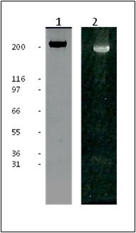
Fibronectin is purified from bovine plasma. Protein purity is determined by scanning densitometry of Coomassie Blue stained protein on a 4-20% polyacrylamide gel. HiLyte Fluor? 488 labeled fibronectin is >80% pure (Figure 1).
The protein is modified to contain covalently linked HiLyte Fluor? 488 at random surface lysines (2). An activated ester of the fluorochrome is used to label the protein. Labeling stoichiometry is determined by spectroscopic measurement of protein and dye concentrations. Final labeling stoichiometry is 1-3 dyes per protein molecule (Figure 2). HiLyte Fluor? 488 labeled fibronectin can be detected using a filter set of 350-450nm excitation and 500-550 nm emission.
Fibronectin runs as individual subunits on SDS-PAGE with an apparent molecular weight of 230 kDa. FNR02 is supplied as an orange lyophilized powder. Each vial of FNR02 contains 20 μg protein
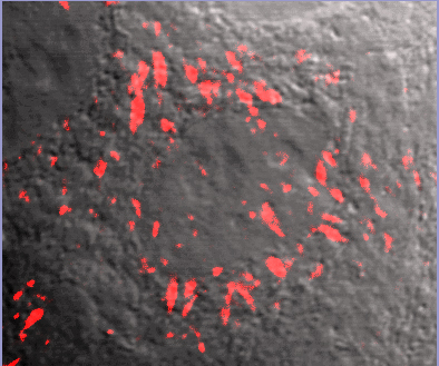
Fluorescent Fibronectin Treated MCF10A cells
Fluorescent fibronectin (Cat. # FNR01) treated MCF10Acells (image kindly provided by A. Varadara and M. Karthykenyan, Univ. S.Carolina,Columbia, SC).
Purity
Purity is determined by scanning densitometry of proteins on SDS-PAGE gels. Samples are >80% pure.


